Search

drnyacoub

Move Better, Perform Better

Category

Sports Pain

Chronic Radiating Neck Pain – Muscles are to blame

The neck muscles aid in the stability and normal functioning of the cervical spine. An impairment in the size of these muscles can lead to the likelihood of radiating neck pain. Evidence has suggested an atrophy of the deep muscles of the neck as a risk factors of neck pain. Radicular neck pain is defined as a pain that stems from the compression or irritation of a nerve root which exists the neck that radiates to the upper extremity.

A recent study has shown there was a significantly higher change in deep neck muscle size between patients who had chronic radiating neck pain and controls. The deep neck muscles for patients were significantly less in size than those without neck pain, which suggests an inefficient muscular system would present with this patient population. A mechanism by which this may occur is through fear avoidance behavior in those with chronic neck pain. As one develops pain in their neck, the fear to move develops and atrophy of muscles becomes prominent thus potentially leading to nerve related syndromes producing pain. Furthermore, a leading cause of nerve injuries to the neck is from cervical disc herniations, and another study has shown an asymmetry between the deep neck muscles sizes in patients who present with cervical disc herniations. Thus, neck pain is associated with impaired muscle function that leads to disuse atrophy and increased neck pain, and the potential risk of neurological pain.

Effective management of disuse atrophy as it relates to neck pain is essential in rehabilitation. The multifidi and longus colli muscles are the deepest tonic and postural paraspinal muscles of the neck, and deserve attention when developing rehabilitation programs. The specific training of atrophied deep cervical muscles may lead to an increase in size and reduce asymmetry thus improving patient outcomes in neck pain.

Two great exercises worth noting to help improve the activation of deep neck muscles are 1) the bird dog exercises and 2) supine chin tuck and lift otherwise known as the craniocervical flexion with or without cervical flexion technique (Shown Below).

Bird Dog Exercise and Supine Chin Tuck Exercise are shown.

Thus, the effective management of your deep neck muscles can help prevent and reduce neck pains associated with neve related discomfort. Working with your Doctor and therapist is essential to having great back health. Always consult your health care professionals before performing any exercise.

Featured post

Non Surgical Management of a Ganglion Cyst

 A dorsal ganglion cysts can both be painful and limiting , and is the most common soft tissue mass found in the hand and wrist. They are thought to be developed from joint stress and degeneration of connective tissue. While studies do show a spontaneous recovery in 8-50% of cases over 2 years, some are eager to seek out non surgical treatments, as surgical treatments have potential recurrence rates of up to 43%!

A recent study that looked at the use of electroacupuncture in the treatment of a dorsal ganglion cyst showed the success of 6 treatment sessions over 4 weeks. Patient initially reported 4/10 pain with painful flexion and limited mobility. After 6 visits patient reported 0/10, full range without pain and a considerable reduction of cyst like appearance in the dorsum of the wrist. In addition, a return to exercise without pain was achieved!

The analgesic effects of acupuncture are greatly documented within clinical control trials. It is evident that acupuncture can aid in increasing endogeouns opioids, serotonin, dynorphins, and specific pain modulation of the spinal cord, as well as nitric oxide release which increases blood circulation. These mechanisms coupled together can be a plausible cause as to why acupuncture is effective in reducing pain and providing a healing environment.

Thus, Electroacupuncture can be a proven conservative method of care to help reduce ganglion cysts, as demonstrated in research today.

References: Wong et al. 2017, JCMA

Author:
Dr. Nourus Yacoub, DC

Lateral Epicondylitis (Elbow Pain)

Lateral Epicondylitis (Elbow Pain)

Lateral elbow pain is one of the most frequent non-traumatic elbow disorders. The most frequent diagnosis is lateral epicondylitis (LE), otherwise referred to as “tennis elbow”. LE is usually caused by the involvement of chronic degeneration of the wrist extensor muscles/tendon as they attach onto the lateral epicondyle of the elbow. There are a small group of forearm and wrist muscles that make up the common extensor tendon of the elbow, and of these muscles the most frequently affected is the extensor carpi radialis brevis. It is reported that repetitive activity may cause the tendon to become inflamed, leading to a tendinosis of the common extensor tendon, which is a progressive degenerative process of the tendon. The picture below demonstrates the injured muscle/tendon fibres involved in lateral epicondylitis. elbow-painGROSS.jpg

How does it all begin?
Tendons stretch easily in response to gradually increasing forces. However, if the stress exceeds the tendon’s capacity to stretching, a small tear called a micro-tear will occur. These micro-tears can multiply and grow over time accelerating the degenerative process, often causing weakening, underuse, weakening at the tendon-bone junction, and poor blood supply (Ahmad et al. 2013).

What are the signs?
It usually starts off with some localized pain, near the lateral aspect of the elbow, radiating down the side of the elbow and outside of the forearm. It can vary between intensity depending on activity, and often is bothersome at sleep (Vaquero-Picado 2016) . It often accompanies with point tender pain, weakness, pain increase with various elbow motions, bony prominences can form along the lateral elbow, and can present with reduced muscle tone and skin turgor. There is no bruising, redness or hotness to the local tissue, and should be revaluated if seen. It is important to also consider other areas of the body that can contribute to elbow pain such as: injuries to the neck that may increase your elbow pain, overuse injuries that are making you use your elbow more (ie, shoulder injury), nerve entrapment, and degenerative disease of bone and cartilage.Shoulder Complex Injuries in Hockey

Recommended Treatment Options
Rehabilitative therapies such as stretching, strengthening, eccentric loading exercises are favorable, as well as activity modifications (Vaquero-Picado 2016). Neuromuscular stabilization techniques of the shoulder, scapula is also important to add to accelerated exercise programing.

Acupuncture has demonstrated very strong evidence in the outcome on short-term treatment and effectiveness (Trinh et al. 2004).

There is some evidence on the short-term use of an elbow strap/brace to reduce pain associated with the tension on the wrist extensor muscles, however, prolonged use is highly cautioned against.

Speaking with your family doctor about alternative to conservative management, that include: corticosteroid injections, PRP injections, medication use.

Most patient’s symptoms resolve with conservative management including manual therapy, activity modifications and exercise. Remember, always try to start your treatment early.

BW Profile Pic MINI

Dr. Nourus Yacoub, DC
Medical Director and Chiropractor
Royal Chiropractic and Sports Injury Clinic

References

Ahmad Z, Siddiqui N, Malik SS, et al. Lateral epicondylitis: a of pathology and management. Bone Joint Journal. 2013; 95-B:1158-1164.

Trinh KV, Phillips S-D, Ho E, Damsma K. Acupuncture for the alleviation of lateral epicondyle pain: a systematic review. Rheumatology (Oxford) 2004;43:1085-1090.
Vaquero-Picado, A., Barco, R. and Antuña, SA. Lateral epicondylitis of the elbow. EFORT Open Rev. 2016 Nov; 1(11): 391–397.


Achilles Tendon Injury

Achilles Tendon Injury

Achilles tendon is located at the back of the leg near the heel. It makes up the confluence of the posterior superficial leg muscles and is regarded as one of the strongest and largest tendon in the human body. Despite its strength, it is frequently subjected to injury and accounts for one of the most common sports injuries, an Achilles tendinopathy. It is important to distinguish between the type of tendon injuries associated with the Achilles.

An Achilles tendinopathy (AT) involves localized pain, swelling near the back of the leg near the heel with impaired physical function. There are two types based on location of injury: the mid tendon, and the distal insertional attachment. An Achilles tendinopathy is usually found in physical activities such as running, sprinting, and jumping, which are common athletic movements in a training program. Some factors to consider which may lead an athlete to higher risk of an Achilles injury include: leg length discrepancy, hyperpronation (flat feet), limited ankle mobility, various health systemic conditions, overloading the tendon, over training, excessive hill training, training on hard surfaces, increasing millage without progressive adaptation, poor shock absorption, and poor choice of footwear. Of course, the latter most risks are potentially avoidable with proper strength and training programs, whereas the former need medical guidance.

When one sustains a Achilles Tendon injury, consistent with a tendinopathy, it is important to consider rehabilitation options and medical management immediately, otherwise if the source of pain persists, the area of degeneration of the tear may worsen overtime, and can lead to an Achilles Tendon rupture (briefly discussed below). While there are no gold standards to treatment, research has suggested these non-operative managements to be helpful: initial rest, walking boot with modified activity, orthotics with specific modifications, graduated heel lifts/wedges that reduce dorsiflexion of the ankle, low level laser therapy, eccentric exercise program, and shockwave therapy, deep friction massage and tendon mobilization. Between a mid tendon and an insertional Achilles tendinopathy, there are specific eccentric exercises that studies have shown to be helpful for an individual’s recovery which will further improve healing and reduce the likelihood of re-injury, or potential rupture if left untreated.

injury-clinic-achilles-tendonitis

In some cases, an Achilles tendon can undergo a traumatic injury in which the tendon can rupture, causing significant deficits. While one would immediately jump to the conclusion that surgery is automatically necessary, advances in non-operative management for acute Achilles tendon ruptures are on the rise. There has been a common consensus in literature to reduce the need to intervene with surgery in Achilles tendon ruptures, where functional outcomes show that there are similar results with pain management and function in the presence of rehabilitation. It appears that early loading has been shown to decrease tendon elongation, improve mechanical properties, and improve functional outcomes. Research has shown that a stepwise progression of tendon loading with exercise, working within tolerable ranges of motion, weaning off mobilizations, and advanced exercise and proprioceptive exercises are all helpful in post injury management.

In all regards, the Achilles tendon, one of the most powerful and strongest tendons in the human body can succumb to injury, and more often in sport related incidents such as running and jumping. It is important to consider rehabilitation options in non-operative management first, as research shows significant results to improving outcome and function when addressed quickly. Next time you, your athlete or loved one complains of pain in the heel/lower part of the leg, be sure to get it checked out!

BW Profile Pic MINI

Dr. Nourus Yacoub, DC
Medical Director and Chiropractor
Royal Chiropractic and Sports Injury Clinic

References

Alfredson H. and Cook J. A treatment algorithm for managing Achilles tendinopathy: new treatment options. British Journal of Sports Medicine. 2007 Apr; 41(4): 211–216.

Shapiro E, Grande D, and Drakos M. Biologics in Achilles tendon healing and repair: a review. Curr Rev Musculoskelet Med (2015) 8:9–17.

Li, H and Hua H. Achilles Tendinopathy: Current Concepts about the Basic Science and Clinical Treatments. BioMed Research International (2016) Volume 2016, 1-9.


AC Joint Separation and the Recovery

Sports injuries are a normal process of athletic development. It is important to understand that injuries always occur and they must be taken care of by health care professionals.images91FQKMWB

Among contact sports, AC joint injuries comprise of about half of all shoulder related injures. The sports most likely to cause AC joint separations are football, soccer, hockey, rugby, and skiing. The most common method of an AC joint separation is falling directly onto the AC joint and/or with the shoulder in an internal and abducted position. In addition, it is important to rule out any concomitant injuries of the glenohumeral joint (shoulder joint) which can simultaneously occur with AC separation injuries. The road to recovery depends on the degree of the injury, a sprain to a full out dislocation warrants good clinical judgement and reasoning to return the athlete back to sport. Here is a brief review of things to consider when recovering from an AC joint separation.

Anatomy and Classification

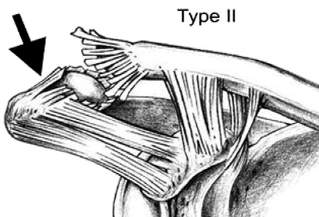
AC ANAtomyTo understand the level of therapy needed to repair an AC separation, it is important to understand the anatomy and the medical classification of an AC joint separation. The AC joint is a small articular joint that links the shoulder girdle to the axial skeleton. It is made up of one small joint (AC), and both the AC ligament and the CC (coracoclavicular) ligaments which attach the shoulder girdle to the clavicle (collar bone).

There are 6 classifications, types 1-3 are the most common, whereas types 4-6 are less common and involve ac-injury.pnga very high degree of impact, and often require surgery consultation. Type 1 involves a sprain of the AC joint with the AC ligament and CC ligaments kept intact. Type 2 involves a subluxation of the AC joint with disruptions of the AC ligament, with a step deformity seen. Type 3 involves both the CC and AC ligaments fully disrupted and a larger step deformity is seen at the AC joint.

Clinical Presentation and Management

Upon initial presentation to the doctor’s office, the patient typically presents supporting their elbow from beneath the injured shoulder using their opposing hand. It is important to recognize shortness of breath as this can lead to a lung injury as well. Abrasions, swelling, bruising may be present on the prominence of the clavicle near the shoulder girdle. Physical exam will include tenderness over the AC joint with minimal range of motion due to discomfort. Orthopedic testing for the AC joint include: cross arm adduction and loading of the AC joint, which will increase pain and localize shoulder pain to the AC joint. These tests are especially useful in patients with type I and II (minor) injuries in which visible or palpable deformity may not be present. Then a shoulder exam will be performed to rule out other injuries to the shoulder capsule. The doctor may decide to send the patient for X-rays of the shoulder and the AC joint.

Treatment Options

AC joint separation management includes acute and sub-acute care with a graduated return to play protocol. Acute and sub-acute management should be focused to relieve the patient from pain, reduce swelling, and mobilization.

Usually the first week of care may require the athlete to wear a cloth sling to prevent movement as the ligaments heal. During active rehabilitation, certain therapeutic modalities can be used to reduce pain and swelling such as electrotherapy and electro-acupuncture, and to help reduce excessive motion the therapist may use athletic taping of the AC joint. In addition, thoracic manipulation can help improve thoracic spine movement, which has been evidenced to help with shoulder mobility.

Usually, after about 3-4 visits (2-3 weeks) of sub-acute management, therapeutic exercises can be added which include light mobility exercise and progressive resistant exercises usually with a frequency of 2 visits per week.

After about 4 weeks of care, a proprioceptive exercise program should be added to improve stability and strength. After about 4-5 weeks of rehabilitation exercises and conservative therapy, pain should subside, and a general exercise program for the athlete should begin which includes upper body strengthening and plyometric exercises. The athlete would be monitored as he/she progresses through their exercise program monthly until they are cleared to return to sport. It is important for the athlete to follow up with their medical provider as they transition week to week, and month to month.

Overall it is important to have comprehensive conservative management when addressing AC joint separations as they can be complex and lead to many limitations as the athlete matures if not properly cleared. The therapist should always evaluate the athlete for contaminant shoulder injuries It is important to at least consult with your medical provider when you suspect an AC injury.

BW Profile Pic MINI

Dr. Nourus Yacoub, DC
Medical Director and Chiropractor
Royal Chiropractic and Sports Injury Clinic

References

Lemos M. The evaluation and treatment of the injured acromioclavicular joint in athletes. Am J Sports Med 1998;26:137-46.

Mazzocca A, Arciero R, Bicos J. Evaluation and treatment of acromioclavicular joint injuries. Am J Sports Med 2007;35: 316-31.

Robb AJ and  Howitt S. Conservative management of a type III acromioclavicular separation: a case report and 10-year follow-up. Journal of Chiropractic Medicine (2011); 10: 261-271.


Importance of a Warm-up

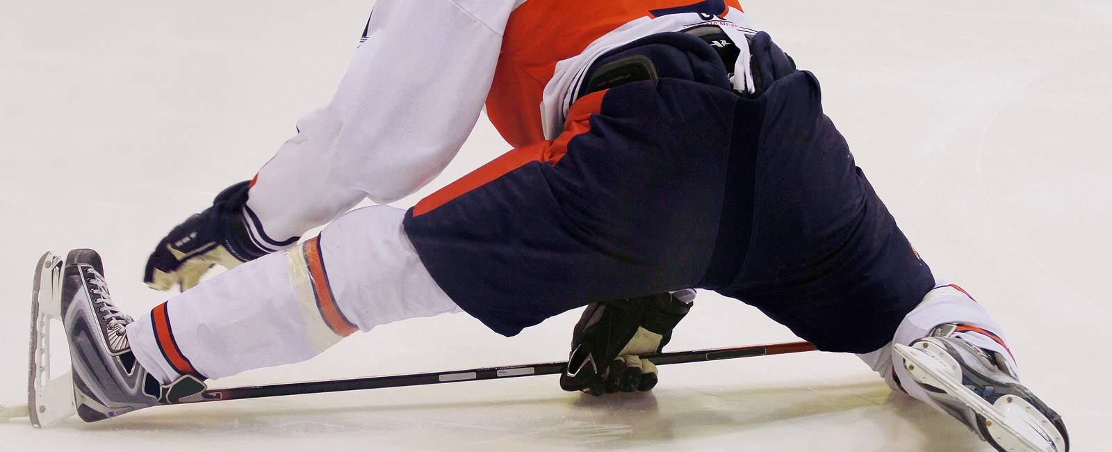
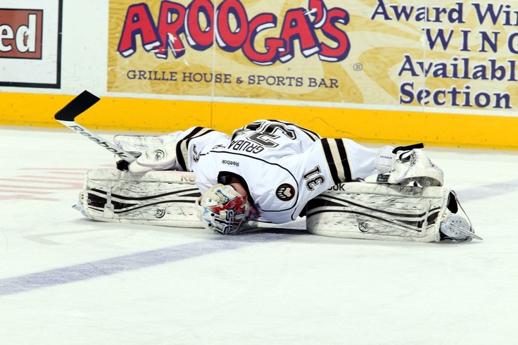
Goalie StretchEvery elite athlete knows that an effective warm-up is an important tool in preventing injury and improving performance. I would like to introduce to you the importance of a warm up, why it is important to have both a static and dynamic component to warm ups and briefly touch on today’s research.

Why Warm up

Warm-ups are important in helping muscle, joints, the heart, and the entire nervous system have a chance to loosen up, and become primed before sport or exercise. As your heart rate increases during a warm up, your blood circulates to internal organs, muscles, tendons and joints, and of course to your brain, which gives your mental game some much added sharpness. It is important to identify sport specific movement patterns and then organize your warmup around these movements. By recognizing these movement patterns, you can then begin to create a warmup specific to your sport which can help activate and prime the joints, muscles and soft tissue involved in sport specific movement.

In private practice, I often run into many young athletes who simply skip the warm up entirely, or don’t adopt an effective routine, which can lead to unwanted injuries. Now that I have illustrated what a warm-up can provide and the specifics of tailoring it to your sport, we must understand the types of warmups there are.

The Dynamic Warm-Up and the Static Warm-Up

When we think of warm-ups, we usually default to a simple hold and stretch type of exercise, that helps loosen some muscles and make us feel good. However, there is much more to a warm-up. There are two types of warm-ups, the dynamic and static warm-up, and they should be used in tandem.

A dynamic warm-up involves actively moving your muscle and joints through cycles of repetition, thus priming movement and preparing muscles for movement in healthy ranges. A static warm-up involves keeping the muscle and joint(s) in motionless position to achieve an increase in flexibility. Let us take the example of an ice hockey goalie who needs to perform splits in a butterfly stance. This athlete’s hip motion needs to have both flexibility and power to properly get in and out of this stance. Training hip flexibility would be achieved using a static warm-up, while a dynamic warm-up can help improve power output and explosive performance of the hip muscles which are needed to achieve proper positioning. While I have illustrated merits to using both methods, it is important to include both, as dynamic warm-ups have been identified in helping reduce any deleterious effects caused by using static warm-ups in isolation. Studies have identified that using only a static warm-up can reduce power output of muscles. So using it just before sport, is not highly recommended, especially in isolation.

What does research tell us

Much of today’s research suggests that static stretching, when used alone, can lead to a reduction in peak power performance and muscle force output. Some researchers suggest that anywhere from 30-90 second hold static stretching can induce these deleterious effects. When a dynamic warm-up is added with a static stretching warm-up, we see much of these deleterious effects reduced. Some research on the use of dynamic warm-ups showed an increase in muscle temperatures, improvements in nerve conduction, an increase in muscle motor unit recruitment, and an increase in the frequency of which fast twitch muscles fire, thus improving force output of the muscle (Layec et al., 2009, Bishop, 2003). Combining a dynamic and static warm-up are both important in helping sport specific movement patterns which can help athletes recover and prevent injuries. Overall incorporating a warm-up with both static and dynamic techniques is extremely important in helping the athlete with flexibility and power output.

A warm-up should include exercises like body weighted lunges, squats, and deadlifts, 20 minutes of cycling, band work, foam rolling, stretches. A common exercises routine that I find a lot of my soccer athletes perform, is the Fifa 11. Try this out, and see if you feel a difference. If you are a follower of my blog, stay tuned to a upcoming program that I am developing with Team Shut Out goalie school.

BW Profile Pic MINI

Dr. Nourus Yacoub, DC
Medical Director and Chiropractor
Royal Chiropractic and Sports Injury Clinic

Resources:

Bishop, D. (2003) Warm up I: potential mechanisms and the effects of passive warm up on exercise performance. Sports Medicine 33, 439-454.

Layec, G., Bringard, A., Le Fur, Y., Vilmen, C., Micallef, J.P., Perrey, S., Cozzone, P.J. and Bendahan, D. (2009) Effects of a prior high-intensity knee-extension exercise on muscle recruitment and energy cost: a combined local and global investigation in humans. Experimental Physiology 94, 704-719.

Samson M, Button DC, Chaouachi A, and Behm, D. Effects of dynamic and static stretching within general and activity specific warm-up protocols. Journal of Sports Science and Medicine (2012) 11, 279-285


Acupuncture, what does it do…

Western medical acupuncture is based on a contemporary understanding of the body’s mechanisms. This practice has earned its place in modern healthcare and has been growing in sports injury management. Western medical acupuncture is used with contemporary medical diagnoses to induce physiological changes in the body, whereas traditional Chinese medical acupuncture is used with diagnoses that arise from understanding the body’s imbalances which are corrected with needles. It is very important to distinguish between the two approaches, as they are used quite differently among practitioners.

There are five mechanisms for understanding how medical acupuncture works in a manual therapy setting, such as in sports injuries and regular sprains and strains.

1) Local Effects

Acupuncture stimulates nerve fibres in the skin and muscle. When needling the skin, it has many sensory nerves that are stimulated and are interconnected within a network. When this network is stimulated, it releases a substance called calcitonin gene-related peptide (CGRP) which causes local blood vessels to expand and increase blood flow. As a result, increases in blood supply can promote local healing of injured tissue.

2) Segmental Analgesia

When an nerve is stimulated, it not only has local effects, but it has effects that reaches the spinal cord and depresses the activity in the region where pain is derived from. It is believed that this process, called, segmental analgesia, inhibits pain using the segmental region of the spinal cord. Acupuncture reduces pain in the segments where the needles are inserted.

3) Extrasegmental Analgesia

After a nerve at the segmental region is stimulated, producing pain relief, the stimulation then travels up to the brainstem, further reducing pain throughout the body. This stimulation is helpful in adding to the reduction of pain at the segment, and else where in the body.

4) Central regulatory effects

As the stimulation of an acupuncture needle can also have effects on structures within the brain. As the midbrain

5) Myofascial trigger points (TrP)

Pain can arise in many different ways, from bones, joints, strained ligaments and tendons, and of course muscles. After heavy overwork, lengthy stretch, awkward posture, muscles can develop small damaged areas that are painful and slow to heal. These small damaged areas begin to manifest into myofascial trigger points (TrP). Usually a TrP can be evident when a taut band of tissues is felt, with a tender spot and reproduced with pressure that can cause referral pain. The patient also complains of biomechanical pain when the area is moved. Acupuncture helps inactivate myofascial trigger points.

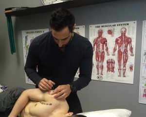
20160602_200537646_iosIn sports injuries, our clinic uses medical acupuncture to the effect of improving tissue healing, improving the neurology of the muscles and tissue, and encouraging an oxygen rich environment for the healing tissue to work in. This often allows the patient/athlete to get back to their sport in a safer manner which is both controlled and monitored, as well as offering a preventative method to future re-injury.

Take home point: Acupuncture is a safe and effective treatment tool for sports injuries, general sprains and strains, and preventative medicine.

BW Profile Pic MINI

Dr. Nourus Yacoub, DC
Medical Director and Chiropractor
Royal Chiropractic and Sports Injury Clinic

References available upon request.


Anterior Knee Pain and OSD

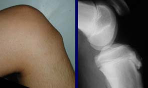
Many children often present in practice complaining about anterior knee pain. One condition that is worth mentioning is Osgood Schlatter disease (OSD). This condition involves an inflammation of the patellar tendon as it inserts on the tibial tuberosity. It is well known to occur in late childhood and often characterised by pain over the bony prominence just below the knee cap (tibial tuberosity). As children are growing, there are growth centers made up of immature bone and cartilage that become mature and ossify into larger bones after skeletal maturity. However, in late childhood, the tibial tuberosity is subject to much stress as activity level increase, and the patellar tendon tugs away from the growth center of the tibial tuberosity leaving inflammation. Pain is usually felt after physical activity, sometimes pain is permanent and steady regardless of the time of the activity. The pain is sometimes followed by a visible inflammation around the patellar attachment of the tibial tuberosity (Figure 1). Activities such as running, climbing up stairs, jumping, bending at the knees can usually exacerbate the symptoms of OSD.

osgood-schlatter_1
Figure 1 – OSD: Anatomcial signs showing palpable growth on tibial tuberosity (left), and Xray findings showing fragmented growth (right).


The first line of defense is resting from pain generating activities, the use of conservative management that targets reduction of pain and swelling, and the use of ice and protective padding. In practice, I tend to use manual muscle release techniques to help reduce the tension along the bony prominence and a combination of electrotherapy and rehabilitation to strengthen muscle groupings. It is important to examine the child’s lower limb biomechanics as poor biomechanical functioning can lead to added complications and poor recovery time.

In some cases, where either conservative management has failed, or the condition has progressed, an x-ray is needed to examine the affected area for bony fragmentation of the tibial tuberosity. And, sometimes an Ultrasound of the soft tissue swelling is important to rule out differential diagnoses. In more extreme cases, a second line of defense involves surgical removal of a bony fragment if pain persists after conservative management. But research tells us that there is no benefit with surgery versus conservative care.

It is also important to consider that there are many other reasons for anterior knee pain such as tumors, infections, muscle and tendon tears, other bony lesions etc.

Take home point: Don’t delay anterior knee pain on a child, get it checked out by a medical health professional.

BW Profile Pic MINI
Dr. Nourus Yacoub, DC
Medical Director and Chiropractor
Royal Chiropractic and Sports Injury Clinic

References
Vaishya R, Azizi A, Agarwal A, et al. (September 13, 2016) Apophysitis of the Tibial Tuberosity (Osgood-Schlatter Disease): A Review. Cureus 8(9): e780


Groin Injuries in Athletes

Hockey-InjuriesGroinGroin injuries occur quite often in sports, and can make a challenging case to treat. Often the symptoms of pain are vague and complicated by multilayered biomechanical deficits. While many athletes are given rest as a form of treatment, this may not prove to be very effective, nor an option for elite athletes who are under pressure to perform.

It is important to be sensitive to groin pain, and it is very important that the area of complaint be evaluated by a health professional familiar with sports related injuries. When dealing with groin pain, it is crucial to understand the different possibilities of injuries. Some injuries that can cause groin pain are groin strains, athletic pubalgia, osteitis pubis, hernias, hip joint arthritis, referral pain, muscle syndromes, and fractures.

Many groin injuries have similar pain patterns, and hence a medical professional must be consulted when groin pain is presented in the athlete. To further understand these injury types, it is important to illustrate that the term “sports hernia”, isn’t a true hernia, as it doesn’t involve a hernia of the abdominal wall like a traditional hernia. Albeit, a sports hernia involves an inguinal disruption, and usually has corresponding MRI evidence of bone marrow edema, capsular ligament disruption and involvement of the enthesis of the hip. Furthermore, a diagnosis of athletic pubalgia is understood to have more of a medial hip pathology involving a disruption of the medial pubic aponeurosis and an adductor tendon pathology, and in some cases the pathology can very much have a disruption of the inguinal ligament, which would fall under the symptoms of a sports hernia. Athletic pubalgia usually presents with chronic pain in the pubic bone, point tenderness along the insertion of the abdominal wall along the pubic tubercle and intermittent weakness of the pelvic wall muscles.


Today most researchers understand the diagnoses of sports hernia as athletic pubalgia, and vice versa. Regardless, of what you would like to call it, the common symptoms of groin pain are understood and the following symptom should be seen upon assessment: deep groin pain/lower abdominal pain, increased pain with exertion, pain over pubic ramus near conjoined tendon, pain with resisted hip flexion at 0, 45, 90 degrees flexion, pain with sit-ups.

Treatment options for inguinal disruptions or athletic pubalgia have been diverse. It is often advisable to begin non-operative treatment immediately, and the program of care should last 8 weeks across 4 phases. The phases are important and overlap one another as the patient progresses. The program aims to reduce swelling and pain, retrain core muscles, hip muscles, and improve global range of motion in the hip and spine, reduce soft tissue restrictions, balance retraining, neurofunctional retraining, and return to sport protocols all within 8 weeks. It is important to prevent the athlete from further damaging the area with over activity and poor biomechanical movements.

Clinical Pearls

Personally, I have seen many groin injuries, athletic pubalgia, sports hernias, hip pointer injuries, and more. They have all been successfully treated at my clinic with my proven methods of treatment. It is important to consider the athlete’s unique injury, as many present with different symptoms, some with MRI evidence, some without. The clinical assessment is my best tool in helping the athlete return to sport, and perform at his preinjury level. I adopt a unique range of treatment that includes medical acupuncture, chiropractic adjustments, soft tissue therapy, exercises and rehabilitation and sport specific rehabilitation.

The most important advice is to get the groin pain checked out. It can save your athletic career!

BW Profile Pic MINI
Dr. Nourus Yacoub, DC
Medical Director and Chiropractor
Royal Chiropractic and Sports Injury Clinic

References
Ellsworth A, et al. 2014. Athletic Pubalgia and Associated Rehabilitation. The International Journal of Sports Physical Therapy, 9(6): 774-784.


Website Powered by WordPress.com.

Up ↑